Mechanics of brain repair
Microglia close brain wounds not by driving neurogenesis, but by pulling on neighbouring cells to contract the injured tissue.
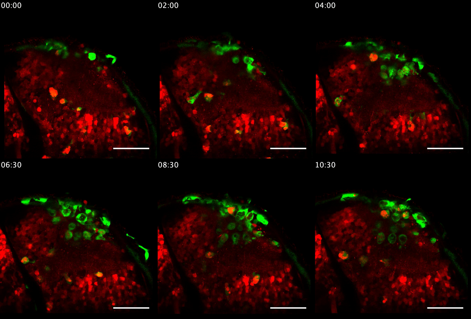
Repairing the central nervous system is a fundamentally physical problem: after a traumatic injury, many cell types — neurons, astrocytes, macrophages, neutrophils — must coordinate to restore the tissue. Working with zebrafish larvae in the lab of Catherina and Thomas Becker, I combined long-term in vivo imaging, controlled laser lesions at cellular resolution, CRISPR genome editing and mathematical modelling to follow this process.
The main finding: microglia are essential for brain repair, not because they trigger neurogenesis, but because they exert local traction forces on neighbouring astrocytes. This drives a large-scale contraction of the tissue that brings surviving neurons back into a configuration where they can re-form a functional visual circuit — challenging the dominant model of repair.
Along the way I developed tools now used by others: a semi-automated laser-lesion protocol adopted by the UK Zebrafish Screening Facility, an observation chamber for long-term imaging on upright microscopes (Wellcome Trust iTPA Translational Innovation Prize, 2020), and a microcontroller-based visual stimulation system.
Key outputs
- El-Daher F., et al. Microglia are essential for tissue contraction in wound closure after brain injury in zebrafish larvae. Life Science Alliance 8(1) (2024). doi:10.26508/lsa.202403052
- El-Daher F., et al. Controlled semi-automated laser-induced injuries for studying spinal cord regeneration in zebrafish larvae. J. Vis. Exp. 177 (2021). doi:10.3791/63259
- El-Daher F., Becker C.G. Neural circuit reorganisation after spinal cord injury in zebrafish. Curr. Opin. Genet. Dev. 64:44–51 (2020). doi:10.1016/j.gde.2020.05.017